
Last Updated on June 18, 2022
When does the aortic semiluna valve close and blood bounces? The aorta is the most elastic tissue in the arterial system, allowing it to expand and contract in response to the flow of blood. This opening allows blood to pass through the valve, but it also distends and closes like a rubber band, causing a secondary pump to inflate arterial pressure.
aortic semilunar valve
When the aortic semilunar valvulus closes and blood bounces, the flow of blood in the aorta oscillates, moving between slightly negative and positive values. The pressure in the aorta typically oscillates between 80 and 120 mm Hg, but this value varies greatly from person to person. Because the aortic semilunar valvulus is so large, pressure waves that have a higher frequency travel faster than those that have lower frequency.
The aortic valve normally consists of three leaflets and a semilunar flap. These valves are attached at the point where the pulmonary artery leaves the ventricles. The aortic valve guards the orifice between the aorta and the pulmonary artery, while the pulmonary valve guards the orifice between the right and left ventricles. These valves have no subvalvular apparatus, making them more similar to the semilunar valves of veins and lymphatic vessels.
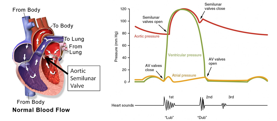
The second heart sound comes from the sudden closure of the semilunar valves at the end of systole. The semilunar valves bulge backwards toward the ventricles and recoils the blood into the arteries. As a result, blood bounces back and forth between the arteries and semilunar valves, creating a resonance that can be heard on the chest wall.
pulmonary semilunar valve
A semilunar valve is a structure made up of connective tissues that sits at the junction of the pulmonary artery and aorta. When the valve is open, blood flows into the aorta and pulmonary arteries in one direction. When it closes, blood returns to the pulmonary arteries in the opposite direction. This explains the second heart sound we hear when the valve closes.
Physiological heart sounds are produced whenever the valves close. These sounds are not directly related to the heart’s contraction, but they are accompanied by distinct sounds that can be heard with a stethoscope. These sounds are produced by the heart’s valves and are recorded by a stethoscope or a digital recorder. Murmurs are generally lower in frequency and occur between 110 and 180 Hz. The second heart sound is a pulsatile sound that originates in the pulmonary artery.
The pulmonary and aortic semilunar valves are pocket-like structures in the heart. These valves protect the aorta and pulmonary artery, respectively, by preventing backflow of blood from the aorta. They act in concert with the AV valves and control blood flow through the aorta. In humans, the semilunar valves open and close as the pressure gradient in the heart changes.
Atrial repolarization
When the aortic semilunar vale closes and the blood bounces off, the heart produces a distinctive sound. This sound is called the “thump” of the heart. In the heart, the aortic semilunar valve is closed when the blood reaches its peak velocity of 30 L/min. In addition, it causes the aorta’s pressure to increase rapidly, far higher than the ventricle’s pressure. So, what does this sound mean?
During rapid ejection, blood flow from the left ventricle to the ascending aorta is the highest. During the decreased-ejection phase, blood flow becomes more gradual and aortic flow starts to decline. The ECG begins at the middle of the P wave and continues with the QRS complex, a prelude to the rise in ventricular pressure.
The aortic valve is one of two semilunar valves in the heart. These valves act in tandem with the AV valves to direct blood flow. When the AV valves are closed, the aortic valve is closed, forcing blood forward. Conversely, when the aortic valve closes and blood bounces, the pulmonary semilunar valve is closed.
In addition to regulating blood flow in the heart, the semilunar valves have several conditions that can affect them. One such condition is stenosis, which occurs when the valves do not fully close. The condition leads to the obstruction of blood flow and, potentially, cardiac failure. Another problem is valve regurgitation, which occurs when the valves are unable to close completely.
Atrial depolarization
In cardiac arrest, the heart stops pumping blood. This uncoordinated, jerky motion is caused by scattered impulses in the myocardium. It is a life-threatening condition, and the only option is to administer a shock, which depolarizes the entire myocardium. If this shock fails to restore normal heart contractions, the patient will likely die.
When the aortic semilunar-valve closes, blood flows from the left ventricle to the ascending aorta. The blood flow from the left ventricle to the aorta increases the most during the rapid ejection phase. The decrease-ejection phase begins when the aortic flow reaches a peak. The jugular venous pulse is included in the third panel of Figure 22-2 for comparison with other events.
During this rapid ejection, the aortic semilunar-valve does not immediately snap shut. The inertia of the blood flow imparts considerable kinetic energy to the blood. The volume of the ventricle decreases less rapidly during phase three-decreased ejection, and the aortic and ventricular pressures fall off. The blood flow rate reaches 70 ml per minute during this phase, and the remaining 50 ml of blood stays in the ventricle.
The pressure wave that travels down the aortic semilunar valvula begins as a single sine wave. As the wave travels down the blood vessel, the peak positions shift backwards. This is called a phase shift, and the magnitude increases with frequency. The higher the frequency, the faster the pressure wave propagates.
Atrial systolic volume
When the aortic semilunar or aortic valve closes and blood bounces, it means that there is a blockage in the flow of blood from the left ventricle into the aorta. The aorta is the largest artery in the human body, and it pumps oxygenated blood to the rest of the body. The aortic valve is one of four chambers in the heart, and it is responsible for pumping blood out to every part of the body. Each chamber has its own sound produced by these four heart valves opening and closing at certain times during the cardiac cycle.
The aortic semilunar valve is the main valve in the heart. Its position in the fetal heart makes it one of the most common to develop stenosis. It has three cusps, each allowing blood to flow in one direction only. The aortic semilunar valve is located between the left ventricle and the aorta. It is also the most likely to be affected by aortic valve disease.
When does the aortic semilunear valve closes and blood bounces? When the aortic semilunar valve closes and the blood bounces, the pressure in the left ventricle is excessively high. Eventually, the pressure in the left ventricle reaches the aorta. The aortic valve opens.
Pulsus paradoxus
When the aortic semilunar heart valve closes, blood bounces off and returns to the heart, a sound is produced in the aorta called a dicrotic notch. It’s important to recognize this sound as the result of an aortic valve occlusion. It can also be the result of a heart condition called pulmonary artery disease.
The aortic semilunar valve is one of four heart valves. It’s the valve that separates the left ventricle from the aorta. It contains three leaflets, known as cusps. Its role is to prevent backflow of blood when the heart is working by preventing the aortic valve from opening. There’s one other valve, called the pulmonary semilunar valve, located between the pulmonary artery and the right ventricle.
When the aortic semilunar vasculature opens, the aortic valve doesn’t immediately snap shut. The blood retains considerable kinetic energy from its rapid backflow. During phase three, when the aortic valve closes and blood bounces, the decrease in ventricular volume slows down. This decrease in ventricular volume makes the aortic and pulmonary pressures fall off. About 70 mL of blood exits the ventricular cavity during this time, while only 50 ml stays in the ventricle.
The aorta and the pulmonary artery were originally one vessel and split into two separate arteries during embryonic development. This valve is a vital piece of the heart, ensuring proper blood flow to the left and right atria. When the aortic semilunar valve closes and blood bounces, the aortic semilunar is not functioning properly.
About The Author

Pat Rowse is a thinker. He loves delving into Twitter to find the latest scholarly debates and then analyzing them from every possible perspective. He's an introvert who really enjoys spending time alone reading about history and influential people. Pat also has a deep love of the internet and all things digital; she considers himself an amateur internet maven. When he's not buried in a book or online, he can be found hardcore analyzing anything and everything that comes his way.