Last Updated on July 27, 2023
Welcome to our article on the fascinating topic of melanocytes! Have you ever wondered where these important cells are located in our bodies? Well, you’ve come to the right place. In this article, we will explore the various aspects of melanocytes, including their function, distribution, and importance for our health. Melanocytes are not only found in our skin, but also in our eyes, hair, and other parts of the body. Understanding their role is crucial for maintaining a healthy body. So, let’s dive in and uncover the mysteries of melanocytes together!
What are melanocytes?
Melanocytes are specialized cells that produce a pigment called melanin. Melanin is responsible for the color of our skin, hair, and eyes. These cells are found in various parts of our body and play a crucial role in protecting our skin from the harmful effects of the sun’s ultraviolet (UV) rays.
Function of melanocytes
- Production of melanin
- Protection against UV radiation
- Determining skin, hair, and eye color
- Regulating skin pigmentation
Distribution of melanocytes in the body
- Skin
- Eyes
- Hair
- Other parts of the body
Melanocytes in the skin
- Located in the epidermis
- Produce melanin to protect against UV radiation
- Determine skin color
Melanocytes in the eyes
- Found in the iris and retina
- Responsible for eye color
- Protect the eyes from UV radiation
Melanocytes in the hair
- Located in the hair follicles
- Produce melanin to give hair its color
- Regulate hair pigmentation
Melanocytes in other parts of the body
- Found in the inner ear, brain, and heart
- Play a role in the development and function of these organs
Importance of melanocytes for health
- Protection against skin cancer
- Regulation of vitamin D production
- Prevention of eye diseases
- Contribution to overall organ health
In conclusion, melanocytes are essential cells that produce melanin and determine our skin, hair, and eye color. They are distributed throughout our body and play a crucial role in protecting us from the harmful effects of UV radiation. Understanding the function and distribution of melanocytes
Function of melanocytes
Melanocytes are specialized cells that play a crucial role in the production of melanin, the pigment responsible for the color of our skin, hair, and eyes. These cells are located in the basal layer of the epidermis, which is the outermost layer of the skin. The main function of melanocytes is to produce and distribute melanin to nearby skin cells. Melanin acts as a natural sunscreen, protecting the skin from harmful ultraviolet (UV) radiation. It also helps to absorb and scatter UV rays, preventing them from penetrating deeper layers of the skin.
In addition to protecting the skin from UV damage, melanocytes also contribute to the process of wound healing. When the skin is injured, melanocytes migrate to the site of the wound and release melanin, which helps to regulate the inflammatory response and promote tissue repair.
Overall, the function of melanocytes is essential for maintaining the health and integrity of the skin. Without these specialized cells, our skin would be more susceptible to sunburn, skin cancer, and other harmful effects of UV radiation.
Distribution of Melanocytes in the Body
Melanocytes, the cells responsible for producing the pigment melanin, are found throughout the body. While they are most commonly associated with the skin, melanocytes can also be found in other parts of the body, including the eyes and hair.
In the skin, melanocytes are located in the basal layer of the epidermis, the outermost layer of the skin. They are scattered throughout this layer, with a higher concentration in areas that are exposed to the sun, such as the face, arms, and legs. This is why these areas tend to have more pigmentation.
In the eyes, melanocytes are found in the iris, the colored part of the eye. The amount of melanin produced by these cells determines the color of the eyes. People with more melanin in their iris tend to have darker eye colors, while those with less melanin have lighter eye colors.
In the hair, melanocytes are located in the hair follicles. These cells produce melanin, which gives hair its color. As we age, the number of melanocytes in the hair follicles decreases, leading to gray or white hair.
Aside from these areas, melanocytes can also be found in other parts of the body, such as the inner ear, brain, and heart. While their function in these areas is not fully understood, it is believed that they play a role in protecting these organs from damage caused by UV radiation.
Overall, the distribution of melanocytes in the body is widespread, highlighting their importance in various physiological processes and their role in maintaining overall health.
5. Melanocytes in the skin
Melanocytes are primarily located in the skin, where they play a crucial role in determining our skin color. Here are some key points about melanocytes in the skin:
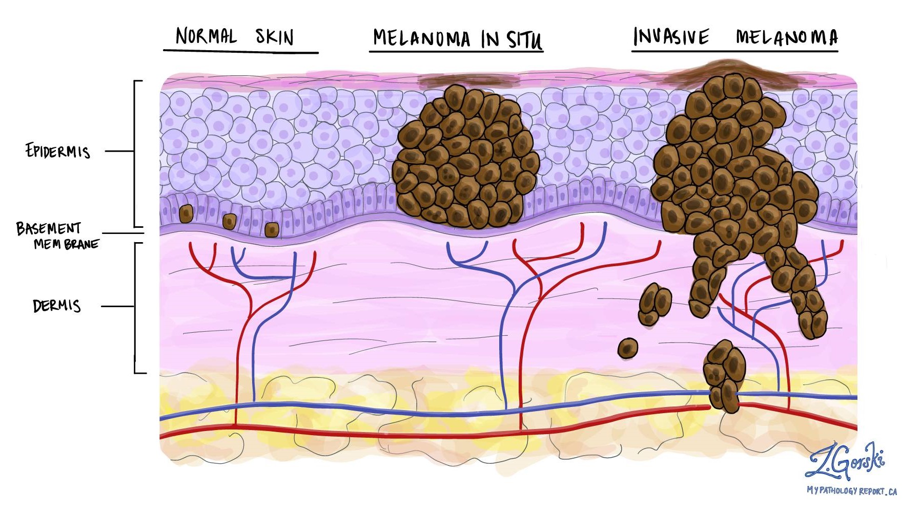
- Melanocytes are found in the basal layer of the epidermis, the outermost layer of the skin.
- They produce a pigment called melanin, which gives color to the skin, hair, and eyes.
- The amount and type of melanin produced by melanocytes determine the color of an individual’s skin.
- People with more melanin have darker skin, while those with less melanin have lighter skin.
- Melanocytes also protect the skin from the harmful effects of ultraviolet (UV) radiation by producing more melanin in response to sun exposure.
- Excessive sun exposure can lead to an overproduction of melanin, resulting in the formation of dark spots or patches on the skin, known as hyperpigmentation.
- On the other hand, a lack of melanin production can lead to a condition called hypopigmentation, characterized by lighter patches of skin.
Overall, melanocytes in the skin are responsible for our skin color and play a vital role in protecting our skin from the damaging effects of the sun.
6. Melanocytes in the eyes
Melanocytes are not only found in the skin, but also in other parts of the body, including the eyes. These specialized cells play a crucial role in the coloration of the iris, which is the colored part of the eye. Here are some key points about melanocytes in the eyes:
- Melanocytes in the eyes are responsible for producing the pigment melanin, which gives color to the iris.
- The amount and distribution of melanin in the iris determine the eye color. People with more melanin have darker eye colors, such as brown or black, while those with less melanin have lighter eye colors, such as blue or green.
- The number of melanocytes in the iris is relatively constant among individuals, but the activity of these cells can vary, leading to different eye colors.
- Changes in the activity of melanocytes in the eyes can occur due to various factors, such as genetics, age, and certain medical conditions.
- Some individuals may have a condition called heterochromia, where they have different colored irises. This occurs when there is a variation in the amount or distribution of melanin in the eyes.
- Overall, melanocytes in the eyes play a significant role in determining eye color and contribute to the uniqueness of each individual’s appearance.
Melanocytes in the Hair
Melanocytes, the pigment-producing cells, are not only found in the skin and eyes but also play a crucial role in determining the color of our hair. These specialized cells are located in the hair follicles, which are tiny structures in the skin that produce hair. The melanocytes produce a pigment called melanin, which gives hair its color.
The amount and type of melanin produced by melanocytes determine the color of our hair. People with darker hair have higher levels of melanin, while those with lighter hair have lower levels. Additionally, the type of melanin produced can vary, resulting in different shades of hair color.
As we age, the number of melanocytes in our hair follicles decreases, leading to gray or white hair. This occurs because the melanocytes produce less melanin or stop producing it altogether. The exact reason for this decline is still not fully understood, but it is believed to be influenced by a combination of genetic and environmental factors.
In conclusion, melanocytes in the hair follicles are responsible for the color of our hair. Understanding the role of these cells can help us better comprehend the mechanisms behind hair pigmentation and the changes that occur as we age.
Melanocytes in other parts of the body
Melanocytes are not only found in the skin, eyes, and hair, but also in other parts of the body. These specialized cells can be found in the inner ear, the brain, and even in the heart. In the inner ear, melanocytes play a crucial role in the production of melanin, which helps protect the delicate structures of the ear from damage caused by loud noises. In the brain, melanocytes are involved in the production of dopamine, a neurotransmitter that is essential for the regulation of movement and mood.
In the heart, melanocytes are responsible for the production of melanin, which helps protect the heart muscle from damage caused by oxidative stress. This is particularly important in individuals with heart disease, as oxidative stress can contribute to the development of cardiovascular complications. Additionally, melanocytes in the heart have been found to play a role in the regulation of blood pressure.
Overall, the presence of melanocytes in these various parts of the body highlights their importance for overall health and well-being.
Importance of Melanocytes for Health
Melanocytes play a crucial role in maintaining our overall health. These specialized cells are responsible for producing melanin, the pigment that gives color to our skin, hair, and eyes. Melanin not only determines our physical appearance but also provides protection against harmful ultraviolet (UV) radiation from the sun.
UV radiation can cause significant damage to our skin cells, leading to sunburn, premature aging, and an increased risk of skin cancer. Melanocytes produce melanin in response to UV exposure, which acts as a natural sunscreen, absorbing and dispersing the harmful rays.
Furthermore, melanocytes also contribute to our immune system. They produce and release various immune-modulating molecules that help regulate the body’s immune response. This is particularly important in the skin, where melanocytes interact with immune cells to defend against infections and other external threats.
Additionally, melanocytes are involved in wound healing and tissue repair. They migrate to the site of injury and release factors that promote the regeneration of damaged tissues. This process is essential for the proper healing of wounds and the restoration of normal skin function.
In summary, melanocytes are not just responsible for our physical appearance, but they also play a vital role in protecting our skin from UV damage, supporting our immune system, and facilitating wound healing. Understanding the importance of melanocytes can help us appreciate the significance of maintaining their health and taking appropriate measures to protect them.
Conclusion: The presence of melanocytes in various parts of the body plays a crucial role in maintaining our overall health and well-being. These specialized cells are responsible for producing the pigment melanin, which gives color to our skin, hair, and eyes. Melanocytes are primarily located in the epidermis of the skin, where they protect us from harmful UV radiation by producing melanin. They are also found in the eyes, specifically in the iris and the choroid, where they help regulate the amount of light entering the eye and protect the delicate structures within. Additionally, melanocytes are present in the hair follicles, where they determine the color of our hair. Furthermore, these cells can be found in other parts of the body, such as the inner ear and the brain, where they have important functions related to hearing and neurological processes. In conclusion, the distribution and function of melanocytes throughout the body highlight their significance for our overall health and well-being.Discover the crucial role of melanocytes in our body and their distribution in the skin, eyes, hair, and more.
About The Author

Alison Sowle is the typical tv guru. With a social media evangelist background, she knows how to get her message out there. However, she's also an introvert at heart and loves nothing more than writing for hours on end. She's a passionate creator who takes great joy in learning about new cultures - especially when it comes to beer!

